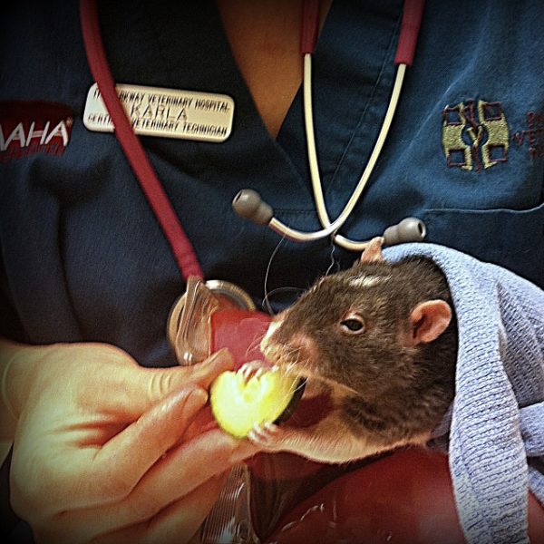
For many years I worked as a receptionist and then as practice manager in various veterinary hospitals. Repeatedly, after a pet’s appointment I’d hear their owner say that the veterinarian “couldn’t figure out what was wrong”. At first I’d sympathize. Then, once I saw that the pet’s owner had declined all diagnostic tests, I’d say to myself, “Of course the doctor couldn’t figure out what was wrong!”
If there’s no blood work, radiographs, or other diagnostic tests performed, doctors are left in the dark. They’re forced to guess what the problem is and they often even prescribe a medication which may or may not be the best choice. Their choice of medication is literally “guesswork”.
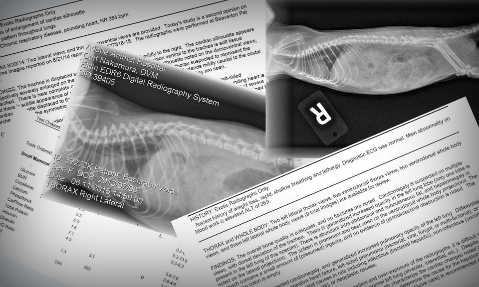
The most common types of diagnostic tests used for pet rats are radiographs (X-rays) and blood work. Sometimes, urinalyses, fecal tests, skin scrapings and cytologies are performed. Less common (but sometimes available) are ultrasounds, electrocardiograms and echocardiograms. All of these diagnostic tools are best performed and interpreted by veterinarians who are knowledgeable and experienced with pet rats. (Not always easy to find.)
The following is a sample of types of labwork and some of the conditions that may be detected: Please note this list is by no means exhaustive. I am simply including the tests with which I am most familiar and which my rats have had over the past 25 years. As long as you have a veterinarian who’s knowledgeable and experienced with pet rats, they’ll be able to tell you which tests will most benefit your rat, adeptly interpret the results and, when applicable, create an optimum treatment plan.
Click on each link (listed alphabetically) to be taken to diagnostic information or scroll down to view:
- Blood Work
- Cytology
- Echocardiogram
- Electrocardiogram
- Fecal Test
- Fine Needle Aspirate
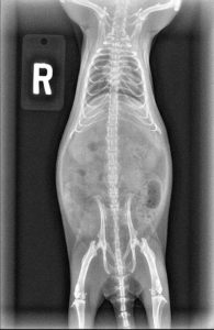
- Radiographs (X-Rays)
- Skin Scraping / Tape Prep
- Ultrasound
- Urinalysis
Blood Work
- What can be detected:
- Elevated liver enzymes
- Kidney disease
- Infection
- Cancer
- Health of adrenal glands
- Immune system status
- Blood sugar levels
- How test is performed:
- Some vets prefer to sedate prior to collecting blood sample to cause less stress for the rat.
- Blood is drawn from a…..
- Jugular vein (neck) – Not done this way frequently since the veins are so tiny
- Saphenous Vein (back leg)
- Tail vein – Most technicians I’ve known prefer this method of collection. I wonder, however, if the rat’s tail is painful afterwards since the skin being punctured to get to the blood vessel is so thick.
- Toe nail – This method of taking blood is not very humane as it definitely is painful afterwards even if rat is sedated during collection.
- Blood is placed in a test tube and spun using a centrifuge before sending to lab
- Interpreting the Results: Lab posts results in veterinary hospital’s online account, usually within twenty-four hours or less. Veterinarian analyzes results and determines any needed treatment plan.
Cytology
- What can be detected
- Types of cells in sample are indicative of what type of mass a pet rat has:
- Cyst
- Abscess
- Benign tumor
- Cancer
- When mass is internal, margins can be evaluated. If mass is cancerous, it’s important to have wide margins so there’s no chance of cancer spreading.
- Types of cells in sample are indicative of what type of mass a pet rat has:
- How test is performed: A very fine needle is inserted into area where fluid or cells exist. This is called a fine needle aspirate. Often with pet rats, it’s an external mass that’s being investigated. Contents of syringe are placed on a glass slide which is stained and then examined under a microscope by a veterinarian and/or submitted to lab for analysis (cytology) by a pathologist. Usually if it’s an external mass, your veterinarian is able to make a determination without sending the sample to lab. If an internal mass has been removed by your veterinarian, it may be suggested the mass be submitted to a lab for analysis. The pathologist will be able to tell what types of cells are in the mass as well as how wide the margins are.
- Interpreting the Results: Lab posts pathologist’s observations and interpretation, usually within twenty-four hours. Veterinarian reviews results and determines any needed treatment plan.

Echocardiogram
- What can be detected:
- Relative sizes of right and left chambers
- Mitrovalve regurgitation
- Size of heart walls
- Shows how blood flows through heart chambers, valves and blood vessels
- How test is performed: In some cases, the rat will be shaved in the area to be examined. A gel is applied to the area which creates a tight bond between the skin and the probe. The gel helps allow the waves to transmit directly to the parts of the rat’s body being imaged. The probe is placed over the rat’s heart. Movement of blood reflects sound waves to the probe or transducer. The transducer sends information to the ultrasound computer. The direction and speed of the blood flowing through the heart and blood vessels can be measured while viewing the patterns shown on the monitor. It’s important to note that not all cardiologists have a small enough probe nor the expertise and confidence to perform an echocardiogram on a rat.
- Interpreting the Results: The echocardiogram is performed by a board-certified veterinary cardiologist who gives a full report of the findings to the primary care veterinarian. In consultation with cardiologist, primary care vet determines what treatment, if any, is needed.
Electrocardiogram (ECG or EKG)
-
- What can be detected:
- Detects heart rate and rhythm, electrical activity of the heart
- Can help identify enlarged and/or overworked heart
- How test is performed:
- Electrodes in the form of small clips are placed on the skin of each limb. Because rats are so small the clips can be uncomfortable since they pinch the skin. Although this test can be stressful, no sedation can be used as it would alter the results. An EKG shows the heart’s electrical activity as a horizontal zigzag on a graph. The spikes and dips denote the waves.
- Interpreting the results: The heart’s electrical activity is recorded as linear spikes and dips which look like a horizontal zigzag running across a graph. If the test is not performed by a veterinary cardiologist, the results will be sent to a cardiologist for analysis. The cardiologist will send a report outlining his or her interpretations of the results to your veterinarian. Your veterinarian will determine what appropriate treatment (if any) is needed.
- What can be detected:
Fecal Test
- What can be detected:
- Intestinal parasites such as roundworms
- Less common intestinal parasites: tapeworms, coccidia
- How test is performed: Sample of feces (as fresh as possible) is brought to vet by owner or collected during an exam. Most veterinarians send sample to a lab for analysis.
- Interpreting the Results: Lab sends report to veterinarian, veterinarian determines (if test is positive) treatment is needed.
- Special Note: I’ve found most vets don’t routinely recommend fecal tests for pet rats. However, I’ve had a few rats with chronic diarrhea. Once the fecal test was run, these rats were placed on appropriate medication for internal parasites found and diarrhea was resolved.
Fine Needle Aspirate
- What can be detected
- Cyst
- Abscess
- Benign tumor
- Cancer
- How test is performed: A very fine needle is inserted into area where fluid or cells exist. Often with pet rats, it’s an external mass that’s being investigated. Contents of syringe are placed on a glass slide which is stained and then examined under a microscope by a veterinarian and/or submitted to lab for analysis (cytology) by a pathologist. Usually if it’s an external mass, your veterinarian will be able to make a determination without sending the sample to lab.
- Interpreting the Results: Most veterinarians are able to detect the type of cells found within an external mass
Radiographs
- What can be detected:
- Respiratory Disease – For example lung consolidation is observed, can indicate respiratory and/or heart disease

- Heart Disease – Enlarged liver and heart as well as lung consolidation can all be indicators of heart disease
- Broken bone(s)
- Internal Masses, Fluid Build-Up – Organs pushed to one side and/or enlarged can indicate a mass and/or fluid build-up
- Intestinal blockage
- Respiratory Disease – For example lung consolidation is observed, can indicate respiratory and/or heart disease
- How test is performed: Some veterinarians will sedate pet rats before taking radiographs. This lessens stress and allows the technicians to optimally position the rat for the radiographs being taken. When appropriate, it’s a good idea to do blood work and radiographs at the same time since having your rat sedated makes it easier for both types of diagnostics.
- There are times when using a dental radiograph machine (normally used for dogs’ and cats’ teeth) is easier and can provide better quality than using the full-body X-ray machine.
- Interpreting the Results: The current best standard of care is for your veterinarian to send out the radiographs taken to be reviewed by a board certified veterinary radiologists who specializes in or has extensive knowledge of exotic pets. The board certified radiologist then sends your vet a report usually within twenty-four hours. Your veterinarian has the option to consult with the radiologist if there are any questions and will determine the best treatment plan for your pet rat based on the radiology report, interpretation and any other test results.
Skin Scraping / Tape Prep
- What can be detected: External parasites
- How test is performed: For a skin scraping, your veterinarian will use a scalpel blade to literally scrape the skin and collect the cells onto a slide. For a tape preparation or cytology, your vet will use a piece of clear adhesive tape applied firmly and repeatedly to the skin and then removed.
- Interpreting the results: The cells that were scraped off by the blade or collected via the clear adhesive tape will be viewed under a microscope. By doing so your veterinarian will possibly see external parasites.
- Special Note: This test is not usually accurate and many veterinarians choose not to perform this test. Even if there are parasites, a skin scraping or tape prep will not always show their presence. Most veterinarians will choose to prescribe a parasiticide based on your rat’s symptoms, history and environment rather than a skin scraping or tape prep.
Ultrasound
- What can be detected:
- Size, shape and location of organs and any masses – if an organ is displaced it can indicate there’s a mass causing the displacement
- Effusion – presence of fluid in lungs and/or in body cavities
- How test is performed: Similar to echocardiogram, probe emits sound waves which are shown as images on a computer monitor. Gel is used to increase efficacy of probe and sometimes rats are shaved to improve contact with skin. Veterinarian may recommend mild sedation for procedure to lessen stress. A board certified ultrasonographer is best equipped to perform the ultrasound, although some exotics vets who have ultrasound machines may possess the knowledge to perform ultrasounds.
- Interpreting the results: If an outside ultrasonagrapher is used, they will submit a report with their findings and interpretations to your rat’s regular veterinarian.
- Special Note: If an outside service is used, it’s important to find an ultrasonographer familiar with exotic pets.
 Urinalysis
- What can be detected:
- Kidney function
- Urine concentration
- Urinary tract or kidney infection
- Diabetes (rare in rats)
- How test is performed: A sample, which needs to be as fresh as possible, is brought in by owner. If sample is collected at night and brought to vet the next morning, it should be refrigerated. Contact your veterinarian for guidance on timing and whether or not to refrigerate. At least 1 ml is generally considered a good amount of urine to collect. Sample is usually submitted to an outside lab for analysis.
- Best ways to collect urine: Keep your rat in a hard plastic carrier (such as one used for cats) without any bedding or litter inside. You can still place food and water inside. If, after several hours, your rat hasn’t urinated inside the carrier, take your rat out and place him/her on a hard, clean surface such as a formica counter top or linoleum on a bathroom floor. They’ll usually urinate once outside the carrier if they’ve been inside for several hours without urinating. On the other hand, many rats will urinate just after they awaken. You may be able to collect urine by placing your rat on a hard surface such as a counter top or bathroom floor as soon as they awaken rather than keeping them for several hours inside a cat carrier. Use a syringe to collect the urine and bring it in to your veterinarian.
- Interpreting the Results: Lab creates report for veterinarian, veterinarian determines what treatment, if any, is needed.
Not mentioned here are fluorescein stains to help detect scratches on eyes (corneal abrasions) or Wood’s lamps (used to see both corneal abrasions as well as skin bacterial and fungal infections such as ringworm) both of which are valuable diagnostic tests for pet rats.
It’s also very important to note that the laboratory used for processing and analyzing tests can make a huge difference. Most veterinary hospitals in the United States use either Idexx or Antech for laboratory analyses. However, there are labs such as Northwest ZooPath which specialize in exotics. Using a lab that’s unfamiliar with normal reference ranges for rats can be dangerous since the results can be misinterpreted.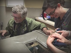
In addition to the lab used, the veterinarian also needs to be familiar with rat physiology and normal reference ranges for laboratory tests. For example, a higher number of mast cells are found in the tissue of rats than are found in cats or dogs. If the lab or veterinarian isn’t knowledgeable about this, a false assumption of mast cell cancer could be made. On radiographs, it’s important to know that the number of lobes in the left and right
lungs is different for rats than it is for dogs or cats. (The left lung of the rat contains one lobe while the right contains four lobes.)
The advantages of rechecks and repeating diagnostic tests cannot be stressed enough. This helps establish patterns and trends helping to determine what is normal for your individual rat (even if it falls outside the normal reference range).
Again, I realize the information here doesn’t include every single possibility when it comes to diagnostic tests for pet rats. My goal, however, is to let you know there are a wide range of options and that with the help of knowledgeable and well-equipped veterinarians, laboratories, pathologists, ultrasonographers, cardiologists and others, much invaluable information can be gained which will help better the lives of our pet rats.
 Urinalysis

